|
An
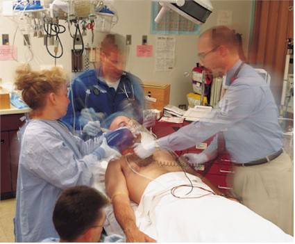
85 year-old man presented to the ED with a two day history
of gradually increasing right eye pain and swelling.
His visual acuity was 20/30 bilaterally and no afferent
pupillary defect was noted. His exam is shown in the
two videos below.


What is your diagnosis and why is this case a bit unusual?
How would you manage this case?
Orbital
Cellulitis
This patient presented with a severe case of orbital
cellulitis, with several interesting features.
Periorbital (preseptal) cellulitis is an infection lying
anterior to the orbital septum. It is usually associated
with swelling of the eyelid, discoloration of the orbital
skin, redness, and warmth. Vision, extraocular movements,
pupillary findings, and optometric examinations are normal.
Orbital cellulitis tends to have similar but more severe
symptoms than preseptal cellulitis. Signs of orbital
cellulitis - in which inflammatory cells and bacteria
invade posterior to the orbital septum to infiltrate the
orbital tissues - include proptosis, decreased ocular
mobility, ocular pain, and tenderness on eye movement.
Limited extraocular movement on the right is evident
on the videos on the preceding page.
Orbital cellulitis is more common in children than in
adults. In one
series of 303 patients with orbital cellulitis, 68% of the
patients were younger than the age of 9 years (1).
A case of orbital cellulitis is indeed unusual in an
85 year-old patient. Moreover,
in adults, orbital cellulitis is typically seen in the
setting of chronic sinusitis.
80-90% of cases in adults are associated with
sinusitis (2). As
is evident on the CT images below, this patient did not
have sinusitis.
Orbital cellulitis generally results from extension of
infection from the periorbital structures, most commonly
from the paranasal sinuses, but also from the face, globe,
and lacrimal sac. Other
etiologies include direct inoculation of the orbit from
trauma or surgery and hematogenous spread from bacteremia.
The etiology in this particular patient was not determined.
Differentiation of periorbital (preseptal) from orbital
cellulitis is an important clinical decision that affects
management and prognosis. If orbital cellulitis is
suspected based on history and examination, a CT scan of
the orbit is indicated to evaluate for subperiosteal or
intraorbital abscess formation.
Images from this patient's CT scan follow.
No abscess was found and, as noted, sinusitis is not
evident. This
patient's CT was notable for considerable edema of the
right orbit anteriorly.
There is mild proptosis of the right eye.
Soft tissue inflammation extends posteriorly
primarily at the medial aspect of the right orbit
|

|

|
Early periorbital (preseptal) cellulitis may be followed on
an outpatient basis for the first 24 to 48 hours of
antibiotic therapy, with daily follow-up to determine
whether resolution is occurring. A broad-spectrum
antistaphylococcal agent provides appropriate coverage.
Treatment for orbital cellulitis includes hospitalization,
intravenous (IV) antibiotics, and occasionally incision and
drainage. Broad-spectrum antibiotic coverage of H.
influenzae, S. aureus, Streptococcus pyogenes, and
anaerobes is indicated. This patient did well with IV
cefuroxime.
If orbital cellulitis progresses, thrombophlebitis may
develop and extend intracranially to cause cavernous sinus
thrombosis. Hallmarks of cavernous sinus thrombosis include
bilateral cranial neuropathy and central neurologic
impairment.
References:
(1) Mawn LA, et al. Preseptal an Orbital Cellulitis Ophthalmology
Clinics of North America 2000; Volume 13, Number 4
(2)Barone ST, et al. Periorbital and orbital cellulitis in
the Haemophilus influenzae vaccine era, J Pediatr
Ophthalmol Strabismus 1997; 34:293
(3) Steinkuller PG, Jones DB: Microbial preseptal and
orbital cellulitis. In: Tasman W, ed: Clinical
Ophthalmology.
Philadelphia
: Lippincott, Williams & Wilkins; 1999: Chapter 25,
1-8, 17-29.
The above case from:

Link
to further Information on:

|


 DISCLAIMER:
This website is designed primarily for use by qualified
physicians and other medical professionals. The
information provided here is for educational and
informational purposes only. It is not guaranteed to be
correct and should NOT be considered as a substitute for
the advice of an appropriately qualified expert. In no way
should the information on this site be considered as
offering advice on patient care decisions or establishment
of a patient-physician relationship.
DISCLAIMER:
This website is designed primarily for use by qualified
physicians and other medical professionals. The
information provided here is for educational and
informational purposes only. It is not guaranteed to be
correct and should NOT be considered as a substitute for
the advice of an appropriately qualified expert. In no way
should the information on this site be considered as
offering advice on patient care decisions or establishment
of a patient-physician relationship.